
The t-tubular system performs a central position in the synchronisation of calcium signalling and excitation-contraction coupling in most striated muscle cells.
Light microscopy has been used for imaging t-tubules for nicely over 100 years and collectively with electron microscopy (EM), has revealed the three-dimensional complexities of the t-system topology inside cardiomyocytes and skeletal muscle fibres from a variety of species.
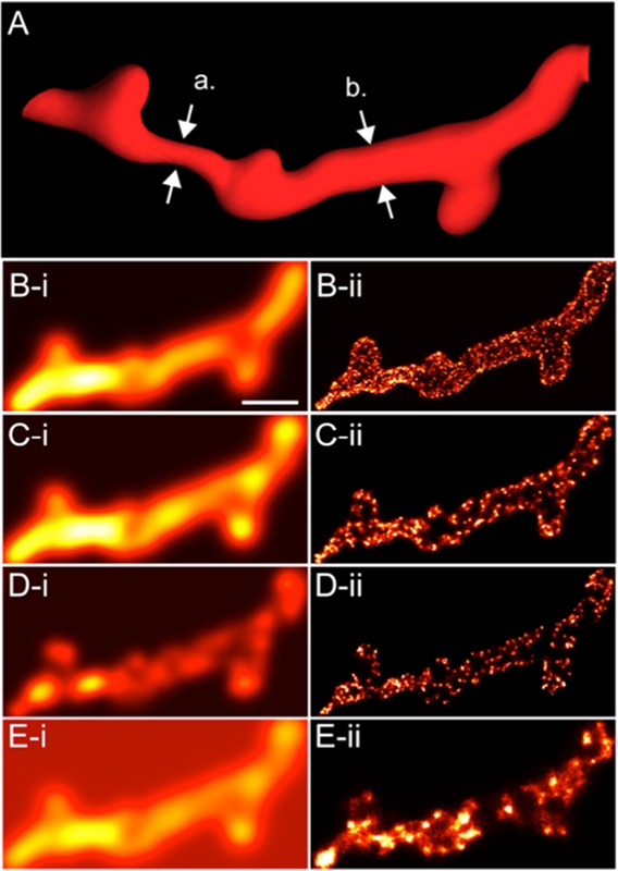
The rising super-resolution single molecule localisation microscopy (SMLM) methods are providing a close to 10-fold enchancment over the decision of typical fluorescence gentle microscopy strategies, with the power to spectrally resolve nanometre scale distributions of a number of molecular targets.
In conjunction with the subsequent technology of electron microscopy, SMLM has allowed the visualisation and quantification of intricate t-tubule morphologies inside massive areas of muscle cells at an unprecedented stage of element. In this paper, we overview current developments in the t-tubule structural biology with the utility of varied microscopy methods.
We define the technical concerns in adapting SMLM to check t-tubules and its potential to additional our understanding of the molecular processes that underlie the sub-micron scale structural alterations noticed in a variety of muscle pathologies.
Nonlinear optical microscopy is a novel software for the evaluation of cutaneous alterations in pseudoxanthoma elasticum.
Pseudoxanthoma elasticum (PXE, OMIM 264800) is a uncommon autosomal recessive dysfunction with ectopic mineralization and fragmentation of elastin fibers. It is brought on by mutations of the ABCC6 gene that results in decreased serum ranges of inorganic pyrophosphate (PPi) anti-mineralization issue.
The incidence of extreme issues amongst PXE sufferers highlights the significance of early analysis in order that immediate multidisciplinary care will be supplied to sufferers. We aimed to look at dermal connective tissue with nonlinear optical (NLO) methods, as collagen emits second-harmonic technology (SHG) sign, whereas elastin will be excited by two-photon excitation fluorescence (TPF).
We carried out molecular genetic evaluation, ophthalmological and cardiovascular evaluation, plasma PPi measurement, typical histopathological examination, and ex vivo SHG and TPF imaging in 5 sufferers with PXE and 5 age- and gender-matched wholesome controls.
Pathological mutations together with one new variant had been discovered in the ABCC6 gene in all PXE sufferers and their plasma PPi stage was considerably decrease in contrast with controls. Degradation and mineralization of elastin fibers and in depth calcium deposition in the mid-dermis was visualized and quantified collectively with the alterations of the collagen construction in PXE.
Our knowledge means that NLO supplies high-resolution imaging of the particular histopathological options of PXE-affected pores and skin. In vivo NLO could also be a promising software in the evaluation of PXE, selling early analysis and follow-up.